Back Of Skull Anatomy : Human Skull Anatomy Hd Stock Images Shutterstock : This view of the skull is dominated by the openings of the orbits and the nasal cavity.
Back Of Skull Anatomy : Human Skull Anatomy Hd Stock Images Shutterstock : This view of the skull is dominated by the openings of the orbits and the nasal cavity.. As these bones grow throughout fetal and childhood development, they begin to fuse together, forming a single skull. Mar 23, 2021 · in all, there are 22 bones comprising the entire skull, excluding the 3 pairs of ossicles located in the inner ear. The entrance to the carotid canal is located on the. These layers of back muscles help to mobilize and stabilize your trunk during your day to day activities. This view of the skull is dominated by the openings of the orbits and the nasal cavity.
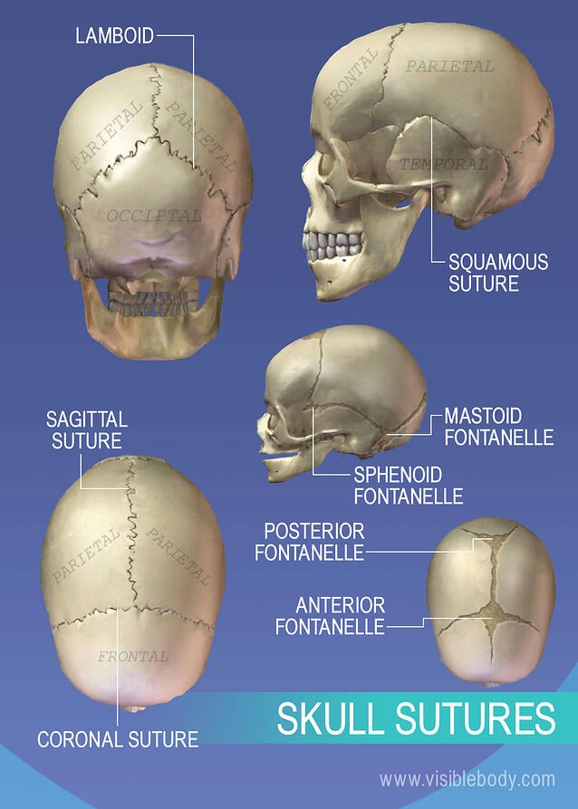
The result is a symmetrically shaped head. Your back consists of three distinct layers of muscles, namely the superficial layer, the intermediate layer, and the deep layer. As these bones grow throughout fetal and childhood development, they begin to fuse together, forming a single skull. The branching pattern of this artery forms readily visible grooves on the internal surface of the skull and these grooves can be traced back to their origin at the foramen spinosum. One suture in the middle of the skull extends from the front of the head to the back.
Human Skull Viewed From The Back Stock Illustration 57701281 Pixta from en.pimg.jp During fetal development, the bones of the skull form within tough, fibrous membranes in a fetus' head. Most of the bones of the skull are held together by firm, immovable fibrous joints called sutures or synarthroses. Also seen are the upper and lower jaws, with their respective teeth (figure 6.17). One suture in the middle of the skull extends from the front of the head to the back. These layers of back muscles help to mobilize and stabilize your trunk during your day to day activities. It is comprised of many bones, formed by intramembranous ossification, which are joined together by sutures (fibrous joints). Jan 12, 2021 · the skull is a bony structure that supports the face and forms a protective cavity for the brain. As these bones grow throughout fetal and childhood development, they begin to fuse together, forming a single skull.
The human crown is made of three layers of the scalp above the skull.
May 19, 2021 · anatomy of back muscles. Your back consists of three distinct layers of muscles, namely the superficial layer, the intermediate layer, and the deep layer. The crown also covers a range of bone sutures, and contains blood vessels and branches of the trigeminal nerve. The human crown is made of three layers of the scalp above the skull. Jan 12, 2021 · the skull is a bony structure that supports the face and forms a protective cavity for the brain. As mentioned, the skull is home to so many structures that the prospect of learning them all can seem very overwhelming. Most of the bones of the skull are held together by firm, immovable fibrous joints called sutures or synarthroses. The major sutures of the skull include the following: It is comprised of many bones, formed by intramembranous ossification, which are joined together by sutures (fibrous joints). May 31, 2021 · skull anatomy diagrams. Also seen are the upper and lower jaws, with their respective teeth (figure 6.17). The result is a symmetrically shaped head. Mar 23, 2021 · in all, there are 22 bones comprising the entire skull, excluding the 3 pairs of ossicles located in the inner ear.
The anatomy of the crown varies between different organisms. May 31, 2021 · skull anatomy diagrams. The result is a symmetrically shaped head. Some sutures extend to the forehead, while others extend to the sides and back of the skull. The major sutures of the skull include the following:
Axial Skeleton Learn Skeleton Anatomy from www.visiblebody.com Most of the bones of the skull are held together by firm, immovable fibrous joints called sutures or synarthroses. The human crown is made of three layers of the scalp above the skull. These layers of back muscles help to mobilize and stabilize your trunk during your day to day activities. During fetal development, the bones of the skull form within tough, fibrous membranes in a fetus' head. May 19, 2021 · anatomy of back muscles. Some sutures extend to the forehead, while others extend to the sides and back of the skull. One suture in the middle of the skull extends from the front of the head to the back. As mentioned, the skull is home to so many structures that the prospect of learning them all can seem very overwhelming.
This view of the skull is dominated by the openings of the orbits and the nasal cavity.
The major sutures of the skull include the following: May 19, 2021 · anatomy of back muscles. These layers of back muscles help to mobilize and stabilize your trunk during your day to day activities. During fetal development, the bones of the skull form within tough, fibrous membranes in a fetus' head. Some sutures extend to the forehead, while others extend to the sides and back of the skull. One suture in the middle of the skull extends from the front of the head to the back. The branching pattern of this artery forms readily visible grooves on the internal surface of the skull and these grooves can be traced back to their origin at the foramen spinosum. The result is a symmetrically shaped head. Jan 12, 2021 · the skull is a bony structure that supports the face and forms a protective cavity for the brain. As these bones grow throughout fetal and childhood development, they begin to fuse together, forming a single skull. The human crown is made of three layers of the scalp above the skull. The bones of the skull are highly irregular. May 31, 2021 · skull anatomy diagrams.
May 19, 2021 · anatomy of back muscles. The bones of the skull are highly irregular. Some sutures extend to the forehead, while others extend to the sides and back of the skull. As these bones grow throughout fetal and childhood development, they begin to fuse together, forming a single skull. Most of the bones of the skull are held together by firm, immovable fibrous joints called sutures or synarthroses.
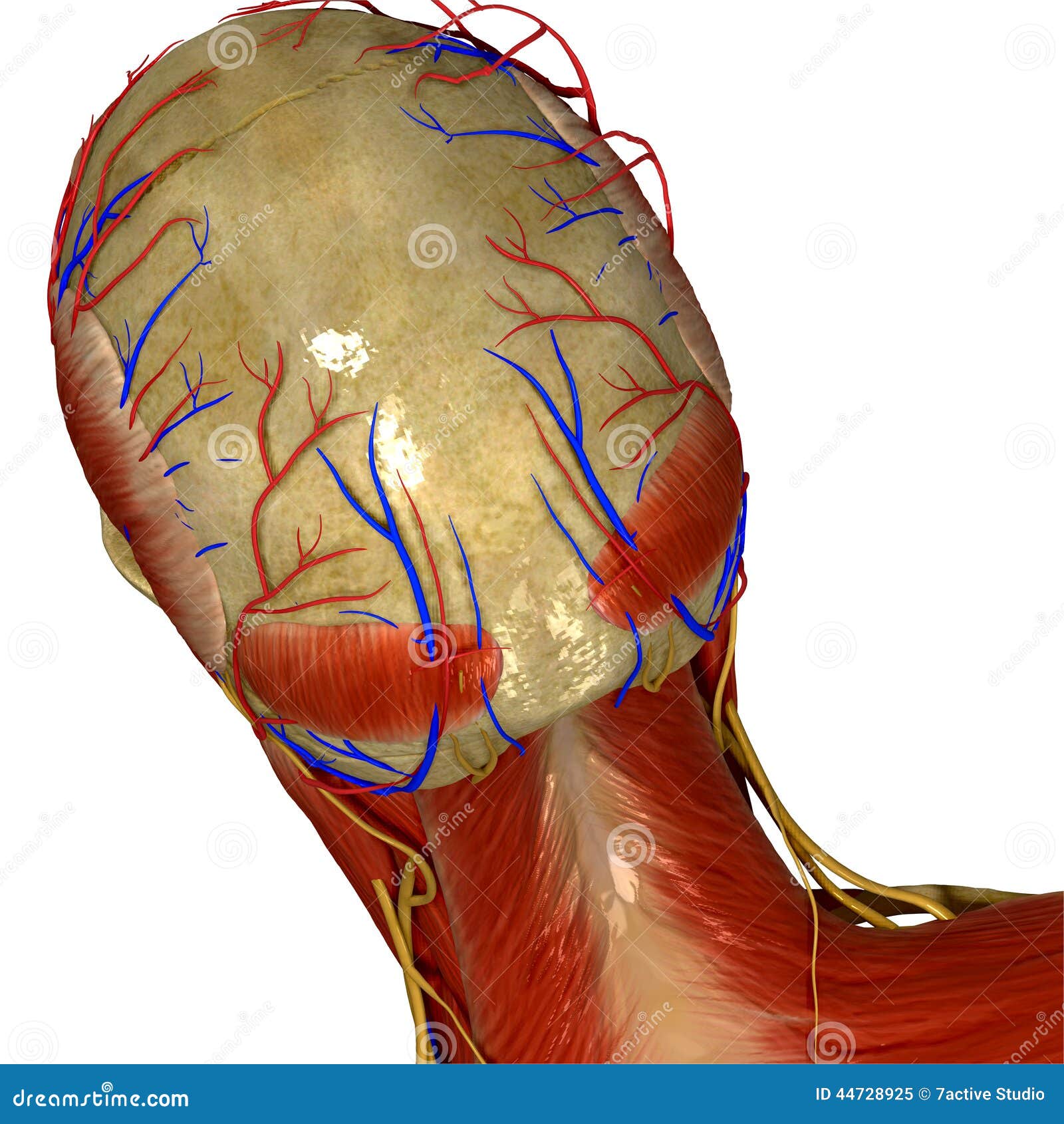
Skull With Muscles And Nerves Back View Illustration 44728925 Megapixl from thumbs.dreamstime.com The branching pattern of this artery forms readily visible grooves on the internal surface of the skull and these grooves can be traced back to their origin at the foramen spinosum. The anatomy of the crown varies between different organisms. During fetal development, the bones of the skull form within tough, fibrous membranes in a fetus' head. The human crown is made of three layers of the scalp above the skull. May 31, 2021 · skull anatomy diagrams. Also seen are the upper and lower jaws, with their respective teeth (figure 6.17). Some sutures extend to the forehead, while others extend to the sides and back of the skull. As mentioned, the skull is home to so many structures that the prospect of learning them all can seem very overwhelming.
It is comprised of many bones, formed by intramembranous ossification, which are joined together by sutures (fibrous joints).
Some sutures extend to the forehead, while others extend to the sides and back of the skull. The anatomy of the crown varies between different organisms. Most of the bones of the skull are held together by firm, immovable fibrous joints called sutures or synarthroses. These layers of back muscles help to mobilize and stabilize your trunk during your day to day activities. The branching pattern of this artery forms readily visible grooves on the internal surface of the skull and these grooves can be traced back to their origin at the foramen spinosum. During fetal development, the bones of the skull form within tough, fibrous membranes in a fetus' head. Mar 23, 2021 · in all, there are 22 bones comprising the entire skull, excluding the 3 pairs of ossicles located in the inner ear. The bones of the skull are highly irregular. As these bones grow throughout fetal and childhood development, they begin to fuse together, forming a single skull. The crown also covers a range of bone sutures, and contains blood vessels and branches of the trigeminal nerve. The result is a symmetrically shaped head. One suture in the middle of the skull extends from the front of the head to the back. This view of the skull is dominated by the openings of the orbits and the nasal cavity.


Posting Komentar
0 Komentar